
Placed, not searched: a protein's shape from its contacts
Can a protein's folded shape be placed directly from a list of which of its parts touch? A measurement on public proteins, with its limits.
Method note
The question
If you know which parts of a protein chain touch each other, can you place the whole chain in its folded shape at once, without searching through the countless ways it could fold?
The result
Told only which parts of a protein chain touch, our engine places the whole chain in its folded shape directly, without searching through possible shapes. No training and no database of known structures enter the method.
On 157 different proteins from the public Protein Data Bank, 46 to 396 amino acids long, the typical placed shape lies 4.26 Å from the measured structure (the median; an ångström, Å, is a ten-millionth of a millimetre). 45% of the proteins come within 4 Å, and 76% within 6 Å.
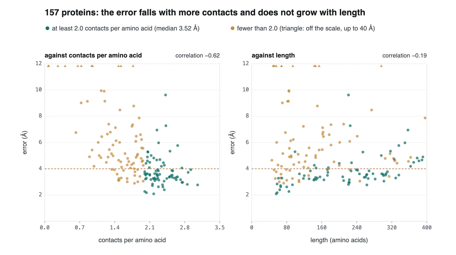
The error does not grow with size. In four size groups of about 40 proteins each, from the shortest chains to the longest, the median error stays between 4.07 and 4.46 Å.
What sets the accuracy is the number of contacts. For the 75 proteins with at least twice as many contacts as amino acids, the median error is 3.52 Å, and 96% come within 6 Å.
Given more than a list of contacts, namely the measured distance between every pair of amino acids closer than 8 Å, our engine rebuilds the same 157 proteins to a median of 1.00 Å; 74% come within 2 Å.
The control
A check that the shape comes from the contacts: where a chain has almost no contacts, it cannot be placed. The two chains with only 4 contacts each end 28 and 40 Å off. Across all 157 proteins, the fewer contacts per amino acid, the larger the error.
The limit
The contacts were read from each protein's measured structure: two amino acids at least five places apart in the chain whose alpha-carbon atoms lie closer than 8 Å. So this measures the placement alone. It is not a prediction from a protein's sequence, and predicting the contacts from a sequence alone is not solved by this work.
The error is the root-mean-square distance between the placed and the measured alpha-carbon atoms, one per amino acid, after the two shapes are laid over each other. The better of the placed shape and its mirror image is counted, because contacts alone do not decide which way the chain turns.
The accuracy is not even: 19 proteins end more than 8 Å off, most of them with few contacts for their length.
The 1.00 Å comes from a second measurement that is given the distances themselves, which is more than knowing which parts touch.
This check comes from the data, not from a planted fault: no run with shuffled or random contacts was made on these 157 proteins.
The engine finds the shape of a protein that already exists; it designs no new protein. It is not compared here with other methods for protein structure.
Data and licence
The data: 157 protein chains from the Protein Data Bank (RCSB PDB), chosen by a fixed, scripted query: X-ray structures at 2.0 Å resolution or better, no two more than 30% alike in sequence, spread evenly over four size groups. From each entry, the longest unbroken stretch of one chain was used.
The data files of the Protein Data Bank are free to use under the CC0 public domain dedication.
Whom to talk to
Questions about this study, or a problem like it in your field: write to us.